马上注册,结交更多好友,享用更多功能,让你轻松玩转社区。
您需要 登录 才可以下载或查看,没有账号?注册
×
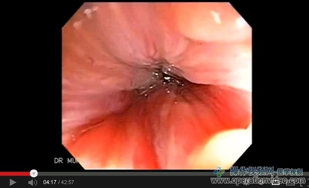
Colonoscopy of Endoscopic Resection of a Rectal Mass
结肠镜检查的内窥镜切除直肠肿块

Published on Apr 23, 2013
Colonoscopy of Endoscopic Resection of a Rectal Mass
Tubulovillous Adenoma with high-grade dysplasia
This is a 75 year-old female, was referred to our endoscopic unit to evaluate a rectal
mass.
Polyps of the colon are mucosal lesions which project into the lumen of the bowel. According to autopsy studies, colonic polyps occur in more than 30% of people over the age of 60. Approximately 70-80% of resected polyps are adenomatous. Adenomatous lesions have a well-documented relationship to colorectal cancer. This adenoma-carcinoma progression represents a significant public health problem, since colorectal cancer is the second leading cause of cancerspecific mortality in the United States. Therefore, appropriate management of colonic polyps may reduce the risk of death from colorectal cancer.
Types of Polyps
There are four types of colonic polyps: adenomatous, hyperplastic, harmartomatous and inflammatory. In addition to these histologic features, polyps are generally described as being either sessile (flat) or pedunculated (having a stalk). Inflammatory and small hyperplastic polyps do not have malignant potential and therefore do not require any further intervention and should not alter surveillance intervals. While most harmotomatous polyps do not have malignant potential, those associated with Peutz-Jeghers syndrome and juvenile polyposis do contain a risk for malignant transformation and therefore require more aggressive intervention and monitoring. Adenomatous polyps are considered precursors for invasive colon and rectal cancer. Histologically these polyps are either villous, tubular or tubulovillous. The risk of malignancy increases with both the size of the polyp and the degree of villous component.
Adenomatous polyps are common neoplastic
lesions of the large intestine. The risk of carcinoma
increases with polyp size. Small polyps are typically
totally embedded for histologic examination, but no
standard method for sampling large, grossly benign
polyps has been established.
Colonic polyps are common specimens encountered in a
pathology practice. It is well established that adenomas
represent precursor lesions to invasive colonic carcinoma.
This is supported by the presence of a spectrum of histologic
changes in adenomas that range from low-grade dysplasia to
frankly invasive carcinoma.
When high-grade dysplasia is
present, there is a strong association with the presence of
invasive carcinoma, and once carcinoma is invasive into
the submucosa, there is a 14% risk for metastasis.
Colonoscopy is the most accurate method for detection of polyps and is the first-line procedure of choice. The sensitivity for detecting polyps by colonoscopy compared to double-contrast barium enema is 94% and 67%, respectively. Although accuracy is operator-dependent, colonoscopy is regarded as the criterion standard.
Cautery snare is recommended for removal of larger polyps. For large sessile polyps, for which the risk of perforation is higher, injection of 1 mL or more of saline into the submucosa directly under the polyp is a useful technique. This lifts the flat polyp away from the muscular layer, creating a stalklike effect. A couple of drops of methylene blue added to the saline also allows the operator to determine if a perforation has occurred in the muscle layer, which would be seen as a break in the layer. Smaller sessile polyps should be removed or biopsied and ablated with hot-biopsy forceps or a minisnare.
After removal of a large (2 cm) sessile polyp or if the possibility exists of incomplete removal of a large adenoma, a follow-up colonoscopy usually should be performed within 3-4 months.
| 

