马上注册,结交更多好友,享用更多功能,让你轻松玩转社区。
您需要 登录 才可以下载或查看,没有账号?注册
×
介绍
左脚踝疼痛和肿胀
患者资料
年龄:45岁
性别:女
X射线
左脚踝
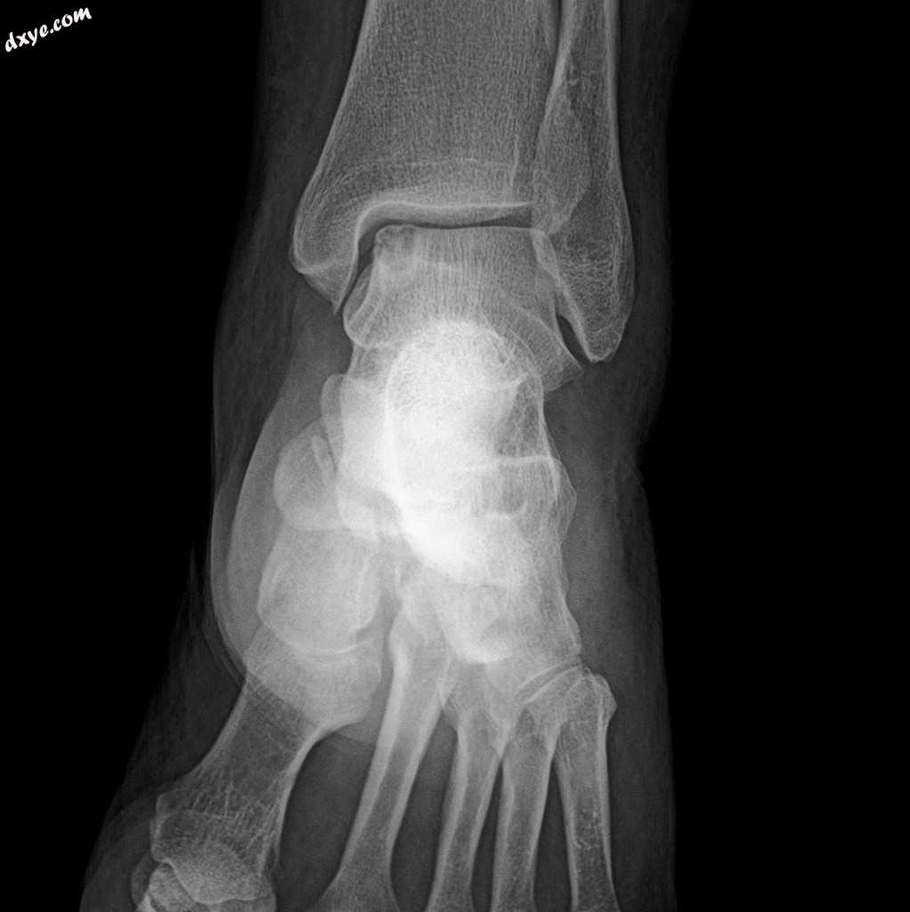
正位
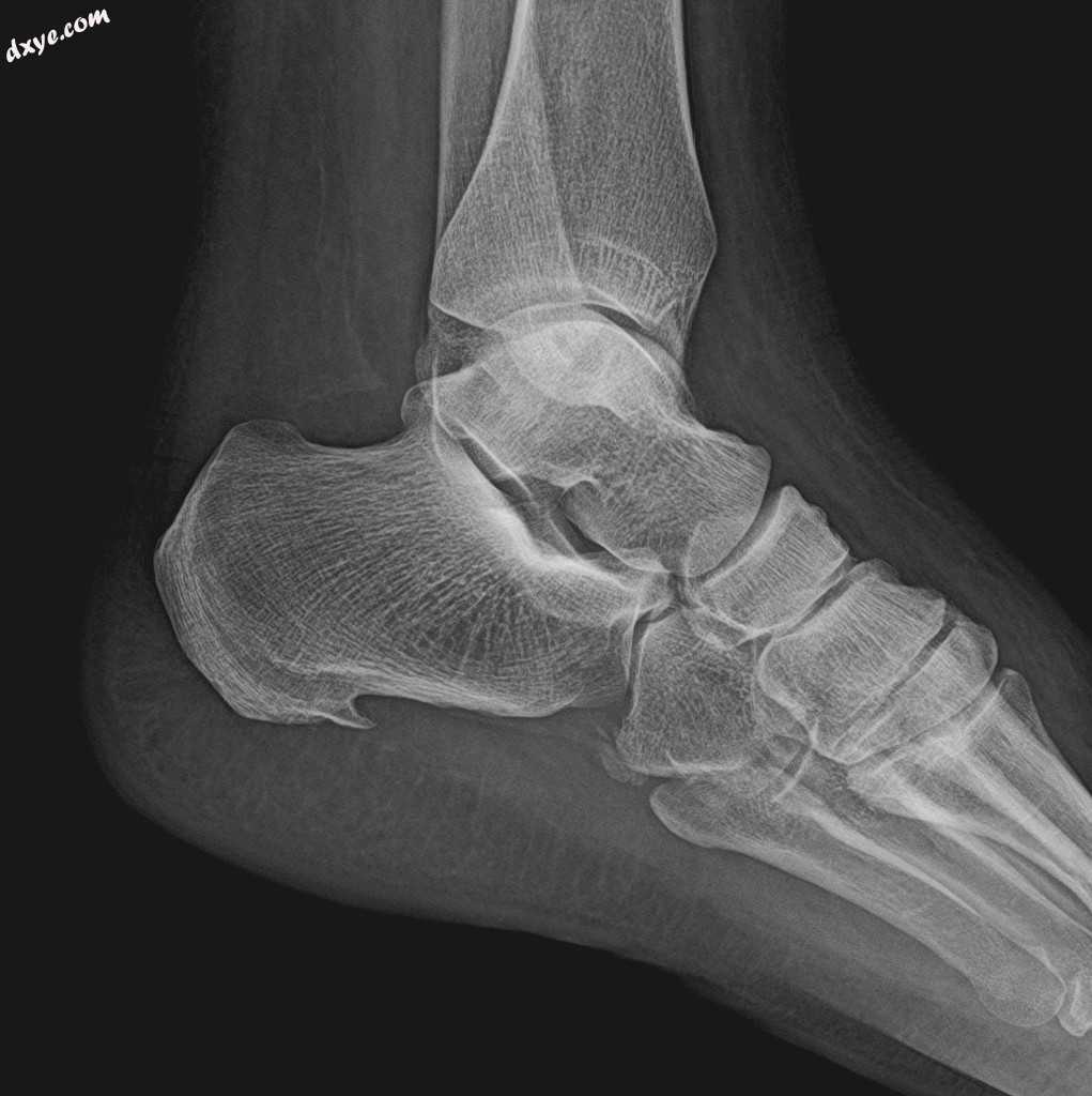
侧位
距骨穹顶的软骨膜下部位可见软骨下圆形透明病变。
注意到跟骨下骨刺。
核磁共振
左脚踝
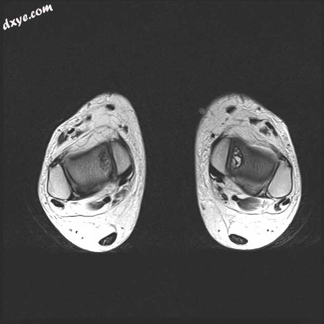
Axial T2
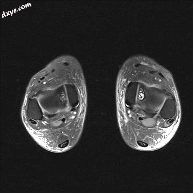
Axial PD fat sat
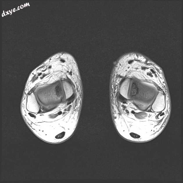
Axial T1
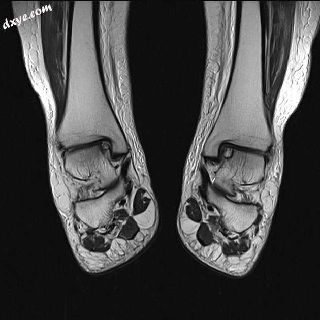
Coronal T2
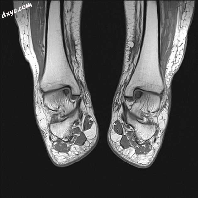
Coronal T1
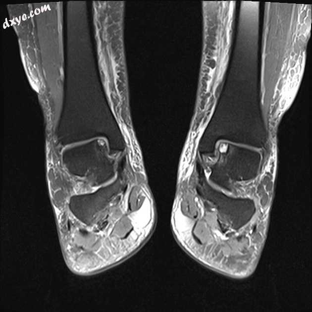
Coronal PD fat sat
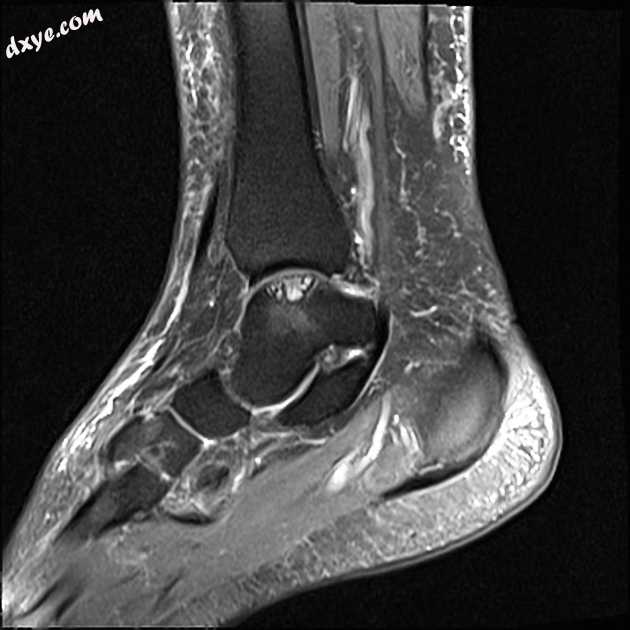
Sagittal PD fat sat
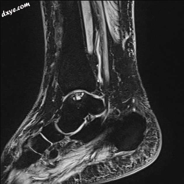
Sagittal WE
右距骨和左距骨的内穹顶的小骨软骨病变,分别为6x10和6x12 mm,周围的骨髓水肿和硬化边缘最小。未见分离的骨碎片。完整的关节表面。
案例讨论
影像学检查显示距骨骨头内侧圆顶的骨软骨损伤。距骨圆顶是骨软骨病变的常见部位,双侧和内侧距骨圆顶病变较常见,占10%。通常,内侧穹顶损伤没有外伤史,与外侧损伤不同。
骨软骨缺损(OCD)或病变(OCL)是关节软骨损伤以及相邻软骨下骨板和软骨下松质骨的损伤的主要病灶区域。
术语
骨软骨缺损是一个广义的术语,描述了关节软骨和软骨下骨中局部间隙的形态变化。它通常与骨软骨损伤/缺损以及儿科人群同义使用。孤立的软骨或软骨下骨病变不被认为是OCD。
请注意,OCD是​​骨软骨缺损和剥离性骨软骨炎(两种密切相关的情况)的常用缩写。
参考资料:
1. Sanders TG, Paruchuri NB, Zlatkin MB. MRI of osteochondral defects of the lateral femoral condyle: incidence and pattern of injury after transient lateral dislocation of the patella. AJR Am J Roentgenol. 2006;187 (5): 1332-7. doi:10.2214/AJR.05.1471 - Pubmed citation
2. Kaplan P. Musculoskeletal MRI. W B Saunders Co. (2001) ISBN:0721690270. Read it at Google Books - Find it at Amazon
3. Sirlin CB, Brossmann J, Boutin RD et-al. Shell osteochondral allografts of the knee: comparison of mr imaging findings and immunologic responses. Radiology. 2001;219 (1): 35-43. Radiology (full text) - Pubmed citation
4. Recht MP, Kramer J. MR imaging of the postoperative knee: a pictorial essay. Radiographics. 22 (4): 765-74. Radiographics (full text) - Pubmed citation
5. Gorbachova T, Melenevsky Y, Cohen M, Cerniglia BW. Osteochondral Lesions of the Knee: Differentiating the Most Common Entities at MRI. (2018) Radiographics : a review publication of the Radiological Society of North America, Inc. 38 (5): 1478-1495. doi:10.1148/rg.2018180044 - Pubmed
6. William Palmer, Laura Bancroft, Fiona Bonar, Jung-Ah Choi, Anne Cotten, James F. Griffith, Philip Robinson, Christian W.A. Pfirrmann. Glossary of terms for musculoskeletal radiology. (2020) Skeletal Radiology. doi:10.1007/s00256-020-03465-1 - Pubmed |


