马上注册,结交更多好友,享用更多功能,让你轻松玩转社区。
您需要 登录 才可以下载或查看,没有账号?注册
×

图-1. 右颞骨。 侧视图。 (From Donaldson JA, Duckert LG, Lambert PR, Rubel EW, eds. Surgical anatomy of the temporal bone, ed 4. New York: Raven Press; 1992.)

图-2. 左颞骨。 后侧视图。 (From Donaldson JA, Duckert LG, Lambert PR, Rubel EW, eds. Surgical anatomy of the temporal bone, ed 4. New York: Raven Press; 1992.)
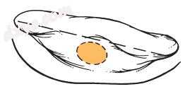
图-3. 耳后切口放置在耳后折痕后约1cm以促进皮肤闭合。 (From Sheehy JL. Surgery of chronic otitis media. In English GE, ed: Otolaryngology. Philadelphia: Lippincott; 1986:1.)

图-4. 最初的切割沿着颞线进行,并与骨管相切。 (From Sheehy JL. Mastoidectomy: the intact canal wall procedure. In Brackmann DE, Shelton C, Arriaga MA, eds: Otologic surgery. Philadelphia: Saunders; 1994.)
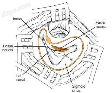
图-5. 面部凹陷显示为三角形。 (From Sheehy JL. Mastoidectomy: the intact canal wall procedure. In Brackmann DE, Shelton C, Arriaga MA, eds: Otologic surgery. Philadelphia: Saunders; 1994.)
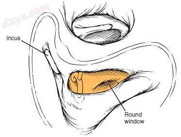
图-6. 面部凹部打开。 (From Sheehy JL. Mastoidectomy: the intact canal wall procedure. In Brackmann DE, Shelton C, Arriaga MA, eds: Otologic surgery. Philadelphia: Saunders; 1994.)
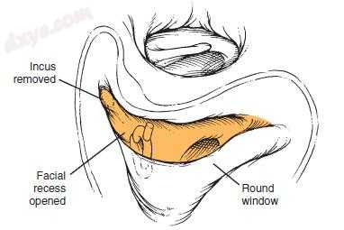
图-7. 通过去除砧骨和颚窝来扩大面部凹陷。 (From Sheehy JL. Mastoidectomy: the intact canal wall procedure. In Brackmann DE, Shelton C, Arriaga MA, eds: Otologic surgery. Philadelphia: Saunders; 1994.)
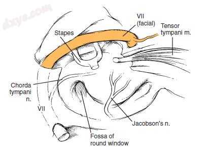
图-8. 面神经鼓室段的过程。 (From Donaldson JA, Duckert LG, Lambert PR, Rubel EW, eds. Surgical anatomy of the temporal bone, ed 4. New York: Raven Press; 1992.)
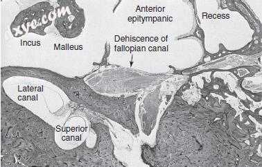
图-9. 前鼓室上空间与开裂面神经。 (From Schuknecht HF. Pathology of the ear, ed 2. Philadelphia: Lea & Febiger; 1993.)

图-10. 管壁式乳突切除术。 CN,颅神经。 (From Brackmann DE, Shelton C, Arriaga MA, eds. Otologic surgery. Philadelphia: Saunders; 1994.)

图-11. 组织切片显示深部窦鼓膜。 EAC,外耳道; N,神经。 (From Schuknecht HF. Pathology of the ear, ed 2. Philadelphia: Lea & Febiger; 1993.)

图-12. A,计算机断层扫描显示继发于慢性中耳炎和由此产生的人工耳蜗瘘(长箭头)的左耳下丘脑细胞系统(短箭头)的侵蚀。 B,磁共振扫描显示钆增强的肉芽组织增强(箭头)。 (From Nadol JB Jr. Revision mastoidectomy. Otolaryngol Clin North Am 2006;39[4]:723-740.)

图-13. 计算机断层扫描显示软组织中的胆脂瘤(箭头)低于左乳突尖端。 在多次修正过程中,胆脂瘤的这个成分被忽略,直到获得该扫描。 (From Nadol JB Jr. Revision mastoidectomy. Otolaryngol Clin North Am 2006;39[4]:723-740.)

图-14. A,轴位计算机断层扫描显示窦和鼓室区的软组织密度(箭头)。 B,与A相同水平的轴向T2加权磁共振扫描显示软组织密度。 C,轴向回声平面扩散加权图像在同一水平显示耀斑“闪耀”效果。 这被放射学解释为胆脂瘤。 (From Evlice A, Tarkan Ö, Kiroğlu M, et al. Detection of recurrent and primary acquired cholesteatoma with echo-planar diffusion-weighted magnetic resonance imaging. J Laryngol Otol 2012;126[7]:670-676.)
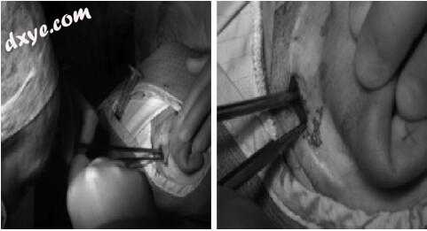
图-15. 外科医生执行45度硬性镜的内窥镜检查。 通过在先前疤痕部位处的耳后刺切口实现通路。 注意这允许双手技术。 (From Barakate M, Bottrill I. Combined approach tympanoplasty for cholesteatoma: impact of middle-ear endoscopy. J Laryngol Otol 2008;122[2]:120-124).

图-16. 逆行乳突切除术技术涉及临时切除上腔静脉壁,伴有逆行乳突切除术。 (Modified from Dornhoffer JL. Retrograde mastoidectomy. Otolaryngol Clin North Am 2006;39[6]:1115-1127.) |


