马上注册,结交更多好友,享用更多功能,让你轻松玩转社区。
您需要 登录 才可以下载或查看,没有账号?注册
×
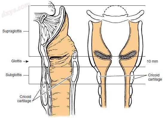
图-1. 根据所涉及的解剖部位分类喉部病变。 (Copyright 2008 by Johns Hopkins University, Art as Applied to Medicine. Modified from Ogura JH, Biller HF. Partial and total laryngectomy and radical neck dissection. In Maloney WH, ed: Otolaryngology, vol 4. New York: Harper & Row; 1971.)
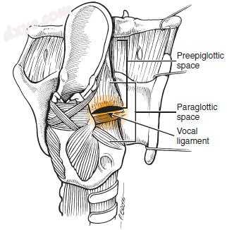
图-2. 显示喉和声门旁间隙合流的喉后倾斜视图。 (From Myers EN, Suen JY, Myers JN, Hanna EYN. Cancer of the head and neck, ed 4. Philadelphia: WB Saunders; 2003.)
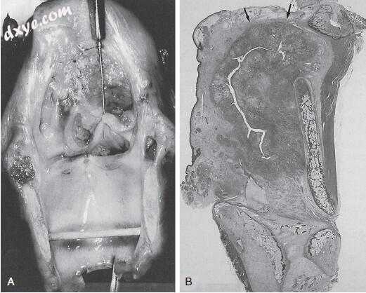
图-3. A,全喉切除标本,伴有深部浸润性声门上型喉癌,起源于假声带。 B,矢状切面显示癌细胞充满了声门旁间隙而没有穿透支气管膜韧带(箭头)。 (From Zeitels SM, Kirchner JA. Hyoepiglottic ligament in supraglottic cancer. Ann Otol Rhinol Laryngol 1995;104:770.)

图-4. 组织切片。 A,会厌喉表面上的癌通过正常的会厌开窗(细箭头)生长到初发性空间。 典型的纤维弹性假包膜在前进癌周围形成(粗箭头)(苏木精 - 伊红;原始放大倍数×40)。 B,会厌鳞状细胞癌的非典型例子位于声门下韧带的下方,没有通过会厌孔进入先声学空间(箭头;苏木精 - 伊红;原始放大倍数×10)。 C,这种鳞状细胞癌在会厌喉表面出现(细箭头),并显示经口腔空洞扩展(粗箭头;苏木精 - 伊红;原始放大倍数×1)在会厌的软骨中通过孔入侵的典型模式。 (From Zeitels SM, Vaughan CW. Endoscopic management of early supraglottic cancer. Ann Otol Rhinol Laryngol 1990;99:951.)

图-5. 喉旁间隙的尺寸位于喉粘膜与其软骨框架之间。 (From Myers EN, Alvi A. Management of carcinoma of the supraglottic larynx: evolution, current concepts, and future trends. Laryngoscope 1996;106:561.)
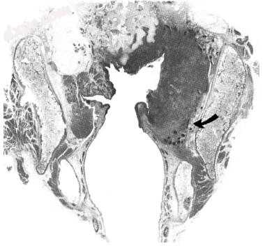
图-6. 此T3声门上癌通过声门旁间隙扩展至声门。 请注意心室底部以下的扩展(箭头),扩大声门旁间隙(苏木精 - 伊红,总冠状面)。 (From Weinstein GS, Laccourreye O, Brasnu D, Tucker J, Montone K. Reconsidering a paradigm: the spread of supraglottic carcinoma to the glottis. Laryngoscope 1995;105:1131.)
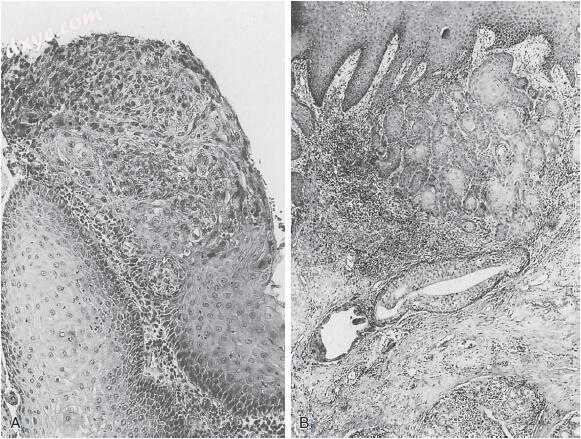
图-7. A,原位癌。 B,微创癌。 在入侵的早期阶段,高分化鳞状细胞的不规则巢浸润固有层并引起炎症反应。 具有鳞状化生的腺体存在于更深的癌细胞附近。 (From Ferlito A, Carbone A, Rinaldo A, et al. “Early” cancer of the larynx: the concept as defined by clinicians, pathologists, and biologists. Ann Otolaryngol 1996;105:245.)
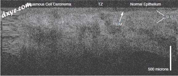
图-8. 放射治疗后复发T2声门鳞状细胞癌的光学相干断层扫描图像。 正常上皮存在于右侧,正常上皮与癌之间的过渡区(TZ)出现在图像的中间。 基底膜(BM)在左侧不可见,对应于浸润性鳞状细胞癌的区域。 SLP,浅层固有层。

图-9. 颈部的六个淋巴结。 (Copyright 2008 by Johns Hopkins University, Art as Applied to Medicine. Modified from Robbins KT, Clayman G, Levine PA, et al. Neck dissection classification update: revisions proposed by the American Head and Neck Society and the American Academy of Otolaryngology–Head and Neck Surgery. Arch Otolaryngol Head Neck Surg 2002;128:751-758.)
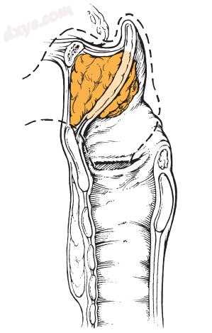
图-10. 开放性声门上型喉切除术的切除概况。 注意包含整个大声空间。 (Copyright 2008 by Johns Hopkins University, Art as Applied to Medicine. Modified from Som ML. Conservation surgery for carcinoma of the supraglottis. J Laryngol Otol 1970;84:657.)

图-11. 原发性声门下淋巴结转移的途径。 沿粘膜下淋巴管扩散至气管旁淋巴结以及沿着下颈静脉链和中颈静脉链。
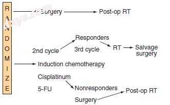
图-12. 来自欧洲癌症研究与治疗组织的治疗算法示意图。 RT,放疗; 5-FU,5-氟尿嘧啶。

图-13. 国家癌症研究所组间喉保存研究示意图。 5-FU,5-氟尿嘧啶; CDDP,顺铂; CR,完整回应; NR,没有回应; PR,部分回应。
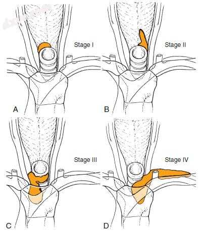
图-14. 造口复发。 A,第一阶段. B,第二阶段。 C,第三阶段。 D,第四阶段。 (From Sisson GA. Ogura memorial lecture: mediastinal dissection. Laryngoscope 1989;99:1264.)
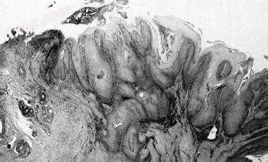
图-15. 一个组织的喉疣状癌显示角化过度、乳头状瘤病,和棒状突(苏木精-伊红;原始放大×25)。 (From Fliss DM, Noble- Topham SE, McLachlin M, et al. Laryngeal verrucous carcinoma: a clinicopathologic study and detection of human papillomavirus using polymerase chain reaction. Laryngoscope 1994;104:147.) |


