马上注册,结交更多好友,享用更多功能,让你轻松玩转社区。
您需要 登录 才可以下载或查看,没有账号?注册
×
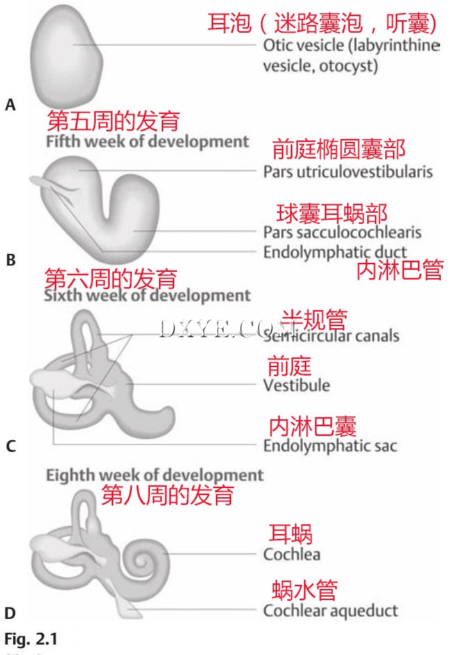
图. 2.1 内耳胚胎学。在胚胎发育的第三周和第四周的皮肤外胚层神经槽之间的上皮增厚的培养形式。 (A) 这种增厚内陷和关闭形成一个独立的囊泡。 (B) 在第五周。 (C) 在第六周,半规管的形式。 (D) 在第七至第九周,耳蜗管形成了囊状囊泡的管状延伸部分并卷曲。 (From Probst R, Grevers G, Iro H. Basic Otorhinolaryngology: A Step-by-Step Learning Guide. Stuttgart/New York: Thieme; 2006:158.)
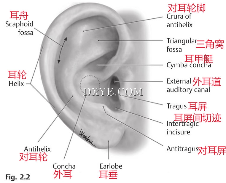
图. 2.2 耳廓解剖学。(From THIEME Atlas of Anatomy, Head and Neuroanatomy, © Thieme 2007, Illustration by Karl Wesker.)
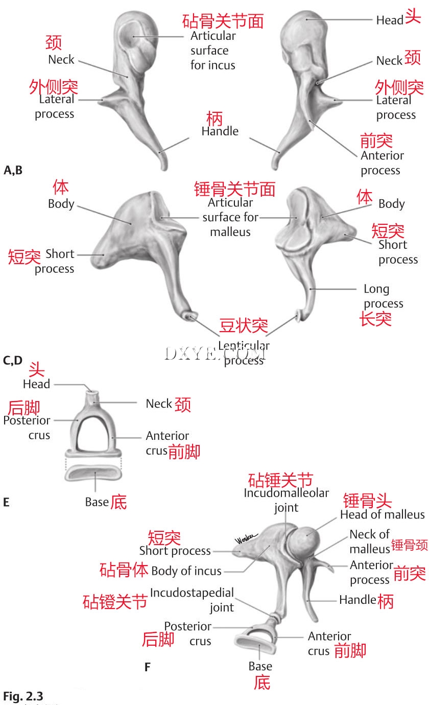
图. 2.3 中耳听小骨。锤骨,后 (A) 和前 (B) 视图。砧骨,内侧 (C) 和前外侧 (D) 视图。 (E) 镫骨,上内侧视图。 (F) 听骨链的内侧面观。 (From THIEME Atlas of Anatomy, Head and Neuroanatomy, © Thieme 2007, Illustration by Karl Wesker.)
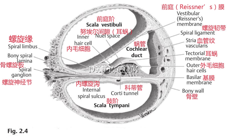
图. 2.4 蜗管。 (From THIEME Atlas of Anatomy, Head and Neuroanatomy, © Thieme 2007, Illustration by Karl Wesker.)
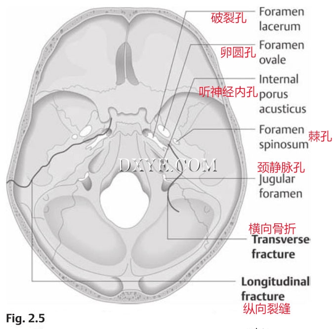
图. 2.5 颞骨骨折:典型的纵骨骨折(左)和横颞骨骨折(右)。(From Probst R, Grevers G, Iro H. Basic Otorhinolaryngology: A Step-by-Step Learning Guide. Stuttgart/New York: Thieme; 2006:303.)
参考:Handbook of Otolaryngology Head and Neck Surgery |


