马上注册,结交更多好友,享用更多功能,让你轻松玩转社区。
您需要 登录 才可以下载或查看,没有账号?注册
×
Laparoscopic resection of a hepatic mucinous cystic neoplasm- A case report
腹腔镜下肝黏液性囊性肿瘤切除术-病例报告
Introduction
We aimed to present a case of hepatic mucinous cystic neoplasm (MCN-H) that was completely resected by laparoscopy.
Presentation of case
引言
我们的目的是提出一个肝粘液性囊性肿瘤病例(mcn-h)是完全腹腔镜切除。
演示案例
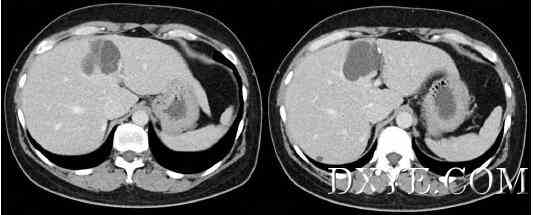
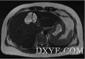
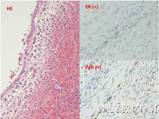
A 47-year-old female exhibited mild elevation of serum liver enzyme levels. Abdominal computed tomography revealed a 45-mm multilocular cystic tumor in segment IV of the liver, along with intermittent border calcification and minimal wall thickness. Magnetic resonance imaging revealed fluid-to-fluid level in the cystic tumor, thereby increasing the suspicion of a mild hemorrhage. The patient underwent laparoscopic liver resection (LLR) with a diagnosis of suspected mucinous cystic neoplasm of the liver. The entire tumor was successfully resected with a laparoscopic approach. The resected specimen was a 4.2 × 3.3 × 2.2-cm cystic tumor. Histological findings revealed mucin-producing singular epithelium and ovarian-like stroma. The tumor was diagnosed as a MCN-H with no malignancy.
一个47岁的女性表现出轻度的血清肝酶水平升高。腹部电脑断层显示的肝段IV 45毫米多房性囊性肿瘤,随着间歇边境钙化和最小壁厚。磁共振成像显示流体在囊性肿瘤中的液位,从而增加了对轻度出血的怀疑。病人接受腹腔镜肝切除术(LLR)与疑似黏液性囊性肿瘤的肝脏。整个肿瘤成功切除与腹腔镜的方法。切除标本4.2×3.3×2.2-cm囊性肿瘤。组织学研究结果显示粘蛋白产生奇异的上皮细胞和卵巢样基质。肿瘤被诊断为MCN-H无恶性肿瘤。
Discussion
讨论
This is the first report in which a MCN-H was completely resected by laparoscopy. MCN-H is rare and is observed in only <5% of liver cystic tumors. MCN-H has been reported to have the malignant potential. And complete resection might be a good treatment option. Along with technical development, LLR has been indicated for benign liver tumors to date. Benign liver tumors are commonly observed in young females. The smaller incisions of the laparoscopic approach might provide cosmetic advantages for patients.
这是第一次报告中的一个mcn-h完全腹腔镜切除。mcn-h是罕见的,只在<5%的肝囊性肿瘤中观察到。mcn-h据报道有恶变可能。完全切除可能是一个很好的治疗选择。随着科技的发展,LLR至今已被证实用于良性肝肿瘤。在年轻女性中常见的良性肝肿瘤。腹腔镜手术的切口较小,为患者提供了美容的优势。
Conclusion
结论
We presented the first case of a MCN-H completely resected by laparoscopy. Benign tumors and tumors with malignant potential might be good indications for a laparoscopic surgery.
我们提出了一个mcn-h完全腹腔镜切除一例。良性肿瘤和潜在恶性可能的肿瘤可能是腹腔镜手术的好适应症。
Keywords: Case report, Laparoscopic liver resection, Liver, Mucinous cystic neoplasm, Ovarian-like stroma
关键词:病例报告,腹腔镜肝切除术,肝,粘液性囊性肿瘤,卵巢样基质
腹腔镜下肝黏液性囊性肿瘤切除术-病例报告

Fig. 1. A multilocular cystic tumor was observed in segment IV of the liver. The tumor was 45 mm in diameter and had septations. CT revealed calcification of the cystic wall and slight wall thickness. There was no mural nodularity or communication to bile ducts.
图1.在肝段IV观察多房性囊性肿瘤。肿瘤直径45毫米,有分隔。CT显示囊壁和微壁厚的钙化。没有壁画结节的通讯或胆管。
腹腔镜下肝黏液性囊性肿瘤切除术-病例报告

Fig. 2. T2-weighted MRI revealed a high intensity on the ventral side and low intensity on the backside of the tumor. Fluid-fluid level was observed on the border, thereby leading to the suspicion that mild hemorrhage may have occurred in the cystic tumor.
图2.T2加强MRI显示高强度的腹侧和低强度的背面上的肿瘤。边界上观察到的液-液平面,从而导致怀疑轻微的出血可能发生在囊性肿瘤。
腹腔镜下肝黏液性囊性肿瘤切除术-病例报告

Fig. 3. The cyst was covered with single layer of epithelial cells (hematoxylin–eosin stain, left). Ovarian-like stroma was observed showing positive immunostaining for both estrogen (upper right) and progesterone receptors (bottom right).
图3.囊肿有上皮细胞单层(hematoxylin–eosin stain,左)。卵巢样基质观察显示雌激素阳性免疫染色(右上)和孕激素受体(右下)。 |
|


