马上注册,结交更多好友,享用更多功能,让你轻松玩转社区。
您需要 登录 才可以下载或查看,没有账号?注册
×
Laparoscopic cholecystectomy using the PINPOINT endoscopic fluorescence imaging system with intraoperative fluorescent imaging- A case report
使用精确内镜荧光成像系统与术中荧光成像的腹腔镜胆囊切除术一例报告

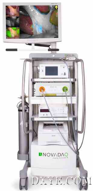
Fig. 1. The PINPOINT endoscopic fluorescence imaging system (Novadaq, Mississauga, ON, Canada; reprinted with permission from Novadaq Technologies Inc.).
使用精确内镜荧光成像系统与术中荧光成像的腹腔镜胆囊切除术一例报告

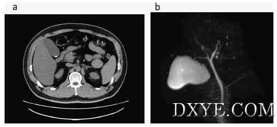
Fig. 2. a. A CT image showing a high absorbance region in the neck of the gallbladder suggesting the presence of a stone. No thickening of the gallbladder wall or increasein the concentration of surrounding fat is evident. Fig. 2b MRI did not indicate an anomaly along the bile duct. The cystic duct branches from the middle portion of the bileduct, and a defect that suggests the presence of a stone in the common bile duct was not observed.
使用精确内镜荧光成像系统与术中荧光成像的腹腔镜胆囊切除术一例报告

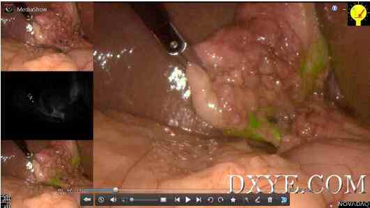
Fig. 3. Laparoscopic images after ICG administration. The gallbladder (arrowhead) and cystic duct (arrow) were imaged in green.
使用精确内镜荧光成像系统与术中荧光成像的腹腔镜胆囊切除术一例报告

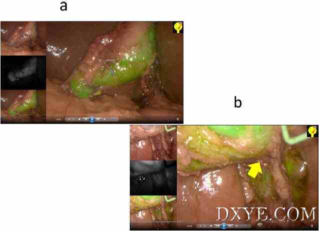
Fig. 4. a. The gallbladder was adequately imaged with ICG fluorescence. Fig. 4b The boundary between the gallbladder and liver was clearly visible (arrow), and the gallbladdercould be detached easily.
| 

