马上注册,结交更多好友,享用更多功能,让你轻松玩转社区。
您需要 登录 才可以下载或查看,没有账号?注册
×
Isolated fallopian tube torsion diagnosed and treated with laparoscopic surgery- A case report
Isolated fallopian tube torsion diagnosed and treated with laparoscopic surgery- A case report

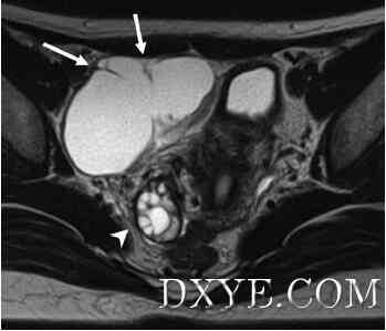
Figure 1. Axial T2-weighted magnetic resonance image showing a cystic lesion in the right pelvis with incomplete wall-like structures (arrows). An intact right ovary can be observed (arrowhead).
Isolated fallopian tube torsion diagnosed and treated with laparoscopic surgery- A case report

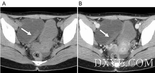
Figure 2. (A) Noncontrast pelvic computed tomographic image showing a cystic lesion in the anterior right pelvis. The partially thickened wall of the cyst shows slightly high attenuation (arrow). (B) Contrast-enhanced pelvic computed tomography scan showing poor enhancement of the cystic wall (arrow).
Isolated fallopian tube torsion diagnosed and treated with laparoscopic surgery- A case report

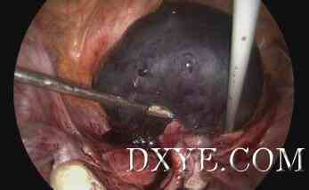
Figure 3. Intraoperative photographic image showing twisted funicular material between the swollen and congested fallopian tube and the healthy fallopian tube.
| 

