- 快捷导航 |


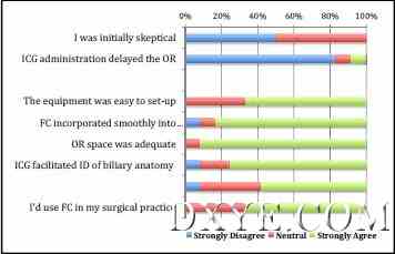
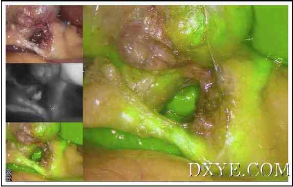
| 荧光造影在腹腔镜胆囊切除术 — 最初的加拿大经验 Fluorescent cholangiography in laparoscopic cholecystectomy- the initial Canadian experience Abstract BACKGROUND: Bile duct injury remains a worrisome complication of laparoscopic cholecystectomy. Indocyanine Green (ICG) fluorescent cholangiography (FC) is a new approach that facilitates real-time intraoperative identification of biliary anatomy. This technology is hoped to improve the safety of dissection within Calot's triangle. 摘要 背景: 胆管损伤仍然是腹腔镜胆囊切除术的一个令人担忧的并发症。Indocyanine Green(ICG)荧光造影(FC)是一种新的方法,有利于胆道解剖,术中实时识别。这项技术有望提高在Calot三角的解剖安全。 METHOD: Demographics, intraoperative details, and subjective surgeon data were recorded for elective cholecystectomy cases involving ICG. Goals were to identify rates of bile duct identification, and assess the perceived benefit of the device. 方法: 人口统计,术中的细节,和主观的外科医生数据记录包括ICG择期胆囊切除术病例。目标是确定胆管的识别率,并评估该设备的感知好处。 RESULTS: ICG was used in 12 biliary cases in Canada. Visualization rates of the cystic and common bile ducts were 100% and 83%, respectively. Also, 83% of surgeons felt that FC incorporated smoothly into the operation. No complications have been related to the technology. 结果: ICG在加拿大12例。胆囊管和胆总管的显示率分别为100%和83%。此外,83%的外科医生认为,FC顺利进入手术。无并发症相关的技术。 CONCLUSIONS: FC allows noninvasive real-time visualization of the extrahepatic biliary tree. This novel technique has received positive feedback in its initial Canadian use and will likely be a durable adjunct for minimally invasive surgery. 结论: FC使肝外胆管树的非侵入性的实时可视化。这种新颖的技术已经收到了积极的反馈,在其初始的加拿大使用,将有可能是一个持久的辅助微创手术。 KEYWORDS: Bile Ducts; Cholangiography; Indocyanine Green; Intraoperative Complications; Laparoscopic Cholecystectomy; Quality Improvement 关键词: 胆管;胆道造影;吲哚青绿;术中并发症;腹腔镜胆囊切除术;质量改进  Figure 1 Survey results from surgeons using FC. Likert scale: 1 to 2, strongly disagree; 3 to 5, neutral; 6 to 7, strongly agree. 图1调查使用FC外科医生的结果。李克特量表:1〜2,坚决不同意;3〜5,中性;6〜7,非常同意。  Figure 2 FC optimizes viewing of the cystohepatic triangle. Clockwise from top left: white light view (standby), NIR overlay (activated display), NIR overlay (standby), subtraction mode (standby). 图2 FC优化观察胆囊三角。顺时针从左上角:白光视图(待机),近红外覆盖(激活显示),近红外叠加(待机),减法模式(待机)。 全文:http://www.dxye.com/thread-24317-1-1.html |