- 快捷导航 |


| 复合顺序旁路使用深静脉搭桥 Composite sequential bypass using profunda vein hitchhike 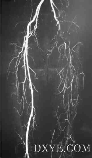 Fig 1. Computed tomography (CT) angiogram of patient 3 showing external iliac occlusion with no reformation in groin. 图1.计算机断层扫描(CT)血管造影的患者3显示髂外动脉闭塞和腹股沟没有改善。 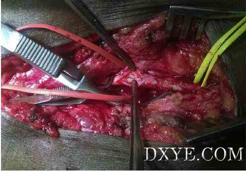 Fig 2. Intraoperative photograph showing fleshy fibrotic material in profunda femoris artery. 图2.术中照片显示在股深动脉的肉质纤维材料。 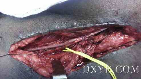 Fig 3. Graft anastomosed to profunda vein and vein graft onto polytetrafluoroethylene graft. Silk tie on the profunda vein proximal to anastomosis. 图3.移植到深静脉和静脉移植到聚四氟乙烯移植吻合。在深静脉近端吻合的丝线结。 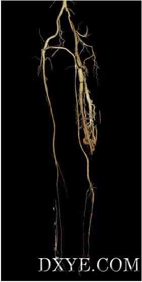 Fig 4. Two-year follow-up angiogram of patient 3 showing functioning graft from right common femoral artery to left anterior tibial artery with left profunda vein as hitchhike with venous collaterals in thigh. 图4。两年随访造影患者3显示功能移植于右股动脉左胫前动脉与左股深静脉与股静脉侧支为搭桥。 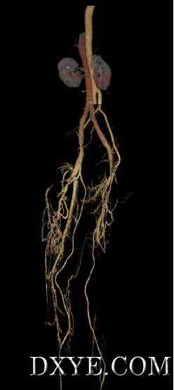 Fig 5. Six-month follow-up computed tomography (CT) angiogram of patient 5 showing patent graft from infrarenal aorta to right peroneal and left anterior tibial arteries with profunda veins on each side as hitchhike points. The angiogram also shows venous collaterals with early filling of the inferior vena cava. 图5.六个月的随访计算机断层扫描(CT)患者5显示专利移植肾下腹主动脉至右腓动脉造影和左胫前动脉、深静脉的每一侧上搭桥问题。血管造影显示静脉侧支与下腔静脉早期充盈。 原文: |